Prostate cancer is one of the most common cancers among men in Israel and worldwide, and is the fourth-highest cause of death from cancer among Israeli males, after lung, colon, and pancreatic cancer. Prostate cancer is characterized by non-typical tissue growth and the creation of a tumor which can penetrate nearby organs or metastasize to distant ones. In recent years, over 2,400 new cases of prostate cancer have been diagnosed annually.
“Due to the high incidence of the disease, emphasis has been placed on early detection, which can enable full recovery as a result of treatment. Sometimes there are no early signs at all, and therefore early diagnosis is extremely important,” notes Dr. Galab Lidawi, a senior physician in the Urology Department at Hillel Yaffe Medical Center.
A biopsy to diagnose or negate a tumor in the prostate gland, is performed today using a transrectal ultrasound. In the past, the doctor used ultrasound to reach the mass using his finger for examination, without knowing exactly where the tumor was located within the prostate gland (“blind biopsy”). With the development of imaging techniques, ultrasound-guided biopsies were taken which sampled areas suspected of having tumors. However, many of the biopsies still gave false information, since when there were small microfocused growths, the biopsy often missed real small but significant areas, meaning up to 35% of serious tumors remained unidentified.

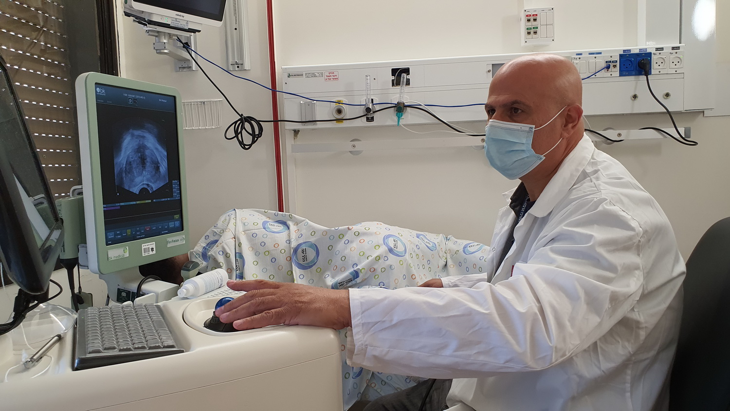
Dr. Lidawi from Hillel Yaffe Medical Center during a combined prostate examination
Recently, a new method for targeted sampling from the prostate, supported by MRI, known as fusion biopsy, has been developed. The test is performed using MRI imaging, and can identify large percentages of significant tumors. Likewise, for the first time the suspicious areas could be identified, measured, and selectively sampled. The test can be carried out under local or general anesthesia.
With this method, the doctor demonstrates the prostate in real time using ultrasound, while watching the pictures of the prostate from the MRI, which were taken earlier and recorded in the device. The MRI and ultrasound images are combined simultaneously using digital technology which enables the places with suspected tumors to be marked precisely. These places are interpreted and marked by a radiologist before the test. Using magnetic resonance imaging, a three-dimensional reconstruction of the prostate is created, and the biopsy is carried out on the reconstructed model in the places marked on the prostate.
In conclusion, notes Dr. Lidawi: “Targeted biopsy using MRI and sonography offers a way of locating and showing areas with suspected cancer with great accuracy. Various studies have demonstrated a success rate of up to 90% for diagnosis or negation of prostate tumors. Combining the pictures provides the urologist with a precise and effective means of diagnosing and treating the disease. In the future, MRI use is likely to become important not just for diagnosis, but also for targeted treatments and follow-up”.