|
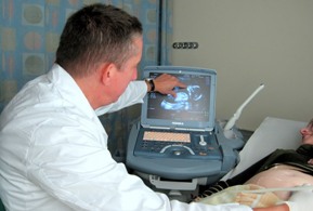
Use of the Doppler ultrasound can help identify fetuses that need to be delivered and significantly lower the risk of intrauterine injury |
Although many factors can affect a fetus's growth potential, the mechanism used to determine the growth rate is not necessarily obvious even today – even if genetic, environmental, nutritional and hormonal factors are known to play a critical role.
Low-weight fetuses had previously been defined as suffering from "retarded intrauterine growth," but since there is no correlation between low weight and (intellectual) delays, the term has justifiably been changed to "intrauterine growth restriction," with anywhere between 3% and 10% of all fetuses being defined as suffering from this type of delay, depending on how it is defined.
When discussing the nature of fetal growth, we should mention the two significant changes that the human body has undergone over the past three million years. The first was the body's transition to an erect position with a significant part of this evolution including adjustment of the bony pelvis. The second change involves the increased volume of our brain from 700 cubic centimeters to 1800 cubic centimeters (vs. the brain of a chimpanzee which grew by only 400 cubic centimeters). As a result, because the fetus's head must pass through the birth canal, these two changes seriously conflict with the need to walk erect – which requires a narrow pelvis – and need to think, which requires a large brain and therefore a large head.
The human body (assisted by evolution) adapted to this conflict in several ways - the first being a shorter pregnancy. If a human being were born with the same level of function as a chimpanzee, the pregnancy would last 17, rather than 9, months.
The second mechanism is stoppage of fetal growth ahead of delivery (around Week 38), something that does not occur in any other animal species.
Fetal growth is divided into three different stages:
The beginning of pregnancy is marked by a massive group of cells (hyperplasia) that is waiting to become organs in the fetus's developing body. Sixteen weeks later, the second stage begins, characterized by production of cells along with growth that already exists. Week 32 of pregnancy is noted mostly for growth of already existing cells (hypertrophia) and the production of new cells decreases. During this period, most of the fetus's fat and glycogen stores form.
In accordance with these stages, the fetus's body grows at a rate of 5 grams per day at the start of the pregnancy, by 15-20 grams per day around Week 24 of pregnancy and by 30-35 grams per day around Week 32 of pregnancy.
Low fetal weight and odds of pregnancy complications and normal growth
In 1963, defined fetal weights for various weeks of the pregnancy were first published. Fetuses with weights defined as being lower than the 10th percentile for the week in pregnancy (i.e. 90% of fetuses were larger) were traditionally defined as suffering from "retarded intrauterine growth" and found to be suffering from unusually high rate of pregnancy complications.
The definitions have since changed and, more importantly, it was found that not all fetuses whose growth was found to be in the lower percentiles were found to be suffering from pathological delay in growth, but some were simply small due to constitutional factors. A key study published in 1992 found that 20%-40% of newborns who were defined as suffering from delayed growth were found to have completely normal growth if several other pieces of information were calculated such as mother's ethnicity, height, weight and even area of residence, since the child's birth weigh changes based on altitude.
As a result, for the purpose of definitions, the use of curves that compare birth weight of infants of similar ethnicities is critical. Israel also has normal growth curves that are based on the Israeli population.
Another study found that within the group of small fetuses, those that suffered from pregnancy complications were primarily the extremely small fetuses (less than the 3rd percentile), and it was therefore argued that the definition must be changed accordingly.
What about fetuses in the normal percentile who began in the upper percentile, but measured in the lower (though still normal) percentile in the ultrasound? In recent years, a great deal of attention has been focused on these types of fetuses. Studies show that even these fetuses who are not in the relatively small percentile, but who are also not maximizing full growth potential have a higher risk of complications at birth.
The expression of small fetus in the post-partum growth process
As previously mentioned, extreme delays in intrauterine growth will be linked to birth complications and occasionally to suboptimal neurological development and increased morbidity in premature infants.
Postnatal growth generally depends on the factor causing the growth delay and in the postnatal environment. Newborns suffering from growth delays attributed to infectious diseases, chromosomal disorders, etc., will generally remain small even after birth. Infants born small due to placental problems will largely catch up with the regular growth rate and maximize their growth potential, depending on optimal growth environment. In other words, a fetus considered small during pregnancy does not necessarily indicate a disrupted growth process unless infectious morbidity occurs or a chromosomal component that indicates problems during pregnancy is detected.
What is the therapeutic approach for suspected small fetus during pregnancy?
If intrauterine restricted growth is suspected during pregnancy, the medical team will try to confirm the diagnosis by ultrasound. Once the diagnosis is confirmed, rare but significant factors will be ruled out such as the presence of fetal defects (identified in scans), chromosomal disorders (suspected or ruled out early in the pregnancy during the nuchal translucency test and several accompanying blood tests), fetal infections (that might be identified in maternal blood tests), maternal illness, etc.
In most cases, when these reasons are ruled out, a certain level of 'placental insufficiency" is suspected. This condition will, in extreme cases, be accompanied by insufficient amniotic fluid, decreased fetal movement and changes in the fetal pulse.
The purpose of treatment, beyond identifying the factors underlying intrauterine restricted growth, is to identify the moment the intrauterine environment becomes dangerous to the fetus, thereby requiring immediate delivery. In order to identify the cases that require delivery at a specific point in time, before changes indicating fetal distress appear, blood flow in placental and fetal blood vessels is measured – use of the Doppler technique. This technology is currently the basis for assessing the condition of fetuses suspected of suffering from delayed growth. The technology is also used to detect other fetal illnesses such as fetal anemia, heart defects, etc.
The ultrasound unit team will sample blood flow from one or more of the placental/fetal blood vessels. Blood flow is generally measured in the umbilical artery (which connects the fetus to the placenta), the middle cerebral artery and the fetal vein. A decrease in flow in these blood vessels is gradual in many cases, indicating that the intrauterine environment is gradually becoming hostile to the fetus – and therefore delivery of the fetus is required.
In conclusion, most fetuses referred to assessment for suspected intrauterine restricted growth will not be found to be significantly smaller when compared with fetuses of the same gender and ethnicity, and taking into account the mother's background. For the fetuses found to be significantly smaller, processing will be carried out to detect the factor or factors behind the delayed growth.
Use of the Doppler ultrasound can facilitate identification of those fetuses that must be delivered and significantly lower the risk of intrauterine injury.
To the Obstetrics, Gynecology and Reproductive Sciences Department>>>


.jpg?BannerID=39 "youtube channel")


